- admin
- June 7, 2026
- 0 Comment
- Highlights | Workshop
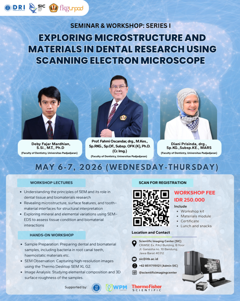
Scientific Imaging Center (SIC), Institut Teknologi Bandung (ITB), officially launched its 2026 workshop series with Seminar & Workshop Series I: Exploring Microstructure and Materials in Dental Research Using Scanning Electron Microscope, held on 6–7 May 2026. Organized in collaboration with the Faculty of Dentistry, Universitas Padjadjaran (FKG Unpad), the workshop highlighted how advanced imaging technologies are contributing to a deeper understanding of dental tissues, biomaterials, and oral health research.
Modern dentistry increasingly relies on microscopic characterization techniques to investigate structures that cannot be observed using conventional optical microscopy. From evaluating enamel and dentin integrity to assessing the performance of restorative materials, researchers require high-resolution imaging tools capable of revealing detailed surface morphology and elemental composition. To address these needs, the workshop focused on the application of Scanning Electron Microscopy (SEM) as a powerful tool for dental and biomaterials research.
The scientific sessions were led by Prof. Fahmi Oscandar, drg., M.Kes., Sp.RKG., Sp.OF., Subsp. OFK (K), Ph.D., Deby Fajar Mardhian, S.Si., M.T., Ph.D., and Diani Prisinda, drg., Sp.KG., Subsp. KE., MARS, all from the Faculty of Dentistry, Universitas Padjadjaran. Through a combination of lectures and demonstrations, the speakers shared their expertise on the role of SEM in investigating dental tissues and supporting the development of innovative biomaterials.
The workshop attracted 21 participants, primarily from Universitas Padjadjaran, Universitas Brawijaya, and Institut Teknologi Bandung. The strong participation from these institutions reflects growing interest in applying advanced microscopy techniques to address challenges in dental science, regenerative medicine, and biomaterials engineering.
A major focus of the workshop was the characterization of dental tissues and restorative materials at the microstructural level. Participants learned how SEM can be used to examine enamel and dentin morphology, evaluate the quality of tooth–restoration interfaces, and assess surface properties of dental biomaterials. The workshop also introduced the use of Energy Dispersive Spectroscopy (SEM-EDS) for elemental analysis, enabling researchers to investigate mineral composition and material interactions that are critical for understanding oral health and treatment outcomes.
Hands-on sessions provided participants with practical experience using the Thermo Scientific Phenom XL G2 Desktop Scanning Electron Microscope. Attendees were guided through sample preparation procedures, imaging workflows, and analytical techniques commonly employed in dental research. The practical training allowed participants to directly observe how SEM data can be used to support scientific investigations and evidence-based clinical research.
Beyond technical skills, the workshop emphasized the broader role of microscopy in advancing dental science. By visualizing structures at the micro- and nanoscale, researchers can gain valuable insights into disease mechanisms, biomaterial performance, tissue regeneration processes, and long-term treatment effectiveness. Such knowledge is essential for developing more durable restorative materials and improving patient care.
Through this specialized program, SIC continues to expand access to advanced imaging technologies while fostering interdisciplinary collaboration between imaging scientists, clinicians, and biomaterials researchers. The workshop demonstrated how electron microscopy serves not only as a research tool but also as a bridge between fundamental science and real-world healthcare applications.
Workshop Highlights
- Event: Seminar & Workshop Series I – Exploring Microstructure and Materials in Dental Research Using Scanning Electron Microscope
- Date: 6–7 May 2026
- Participants: 21 attendees
- Main Participating Institutions: Universitas Padjadjaran, Universitas Brawijaya, and Institut Teknologi Bandung
- Speakers: Prof. Fahmi Oscandar, Deby Fajar Mardhian, Ph.D., and Diani Prisinda, drg., Sp.KG., Subsp. KE., MARS
- Instrument: Thermo Scientific Phenom XL G2 Desktop SEM
- Research Applications: Dental tissue characterization, biomaterial evaluation, enamel and dentin analysis, tooth–restoration interface assessment, and elemental analysis using SEM-EDS
- Outcome: Strengthened understanding of how advanced microscopy can support innovation and evidence-based research in dentistry
By focusing on the intersection of dental science, biomaterials, and electron microscopy, the workshop set the stage for SIC’s 2026 training activities while highlighting the growing role of advanced imaging technologies in improving oral healthcare research.