
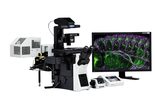
The FLUOVIEW FV1200 combines spectral confocal imaging with advanced fluorescence detection to provide high-quality, quantitative imaging for biological research. Supporting multidimensional acquisition, flexible spectral detection, and powerful image analysis, the system is suitable for both fixed and live-cell imaging. An optional fourth confocal detector increases detection efficiency, enabling reliable imaging of low-intensity fluorescence signals.
About
The Olympus FV1200-SPD is a spectral confocal laser scanning microscope designed for high-resolution fluorescence imaging and quantitative biological research. Equipped with spectral detection, flexible laser control, and an advanced motorized IX83 inverted microscope with Z Drift Compensation (ZDC), the system supports multidimensional imaging including XYZ, time-lapse, and spectral (Lambda) acquisition for both fixed and live-cell applications.
Features
- Spectral confocal imaging with three high-sensitivity PMT detectors
- Flexible spectral detection with 2 nm spectral resolution
- Multi-dimensional imaging (X, Y, Z, Time, Lambda)
- Kalman noise reduction filtering
- Quantitative fluorescence analysis including intensity, area, perimeter, and colocalization
- Advanced 3D visualization with free rotation, volume rendering, stereo view, and animation
- Fully motorized IX83 inverted microscope with Z Drift Compensation (ZDC) for long-term imaging stability
Laser Sources
- 405 nm Laser Diode : DAPI
- 473 nm Laser Diode : FITC, GFP, Acridine Orange, Fluo-3, Fluo-4
- 559 nm Laser Diode : Rhodamine
Scanning System
- Two ultra-high sensitivity PMT spectral detectors
- Scan speed: up to 16 frames/second
Image Analysis
- Image Analysis
- Fluorescence intensity measurement
- Area and perimeter analysis
- Colocalization analysis
- Quantitative fluorescence measurements
- Time-lapse analysis
- Interactive 3D visualization and volume rendering.